A soft marker on ultrasound is a small difference in how the fetus looks on a prenatal scan that is not a defect, does not affect how an organ works, and usually disappears on its own before birth. It is an observation your sonographer records so your clinician can weigh it alongside your age, screening results and the rest of your pregnancy. This guide explains the common findings, what usually happens next, and where reassurance comes from.
This is general health information, not a diagnosis. Only your obstetric clinician, midwife or maternal-fetal medicine specialist can interpret a finding on your own scan.
Table of Contents
- What Does a Soft Marker on Ultrasound Mean?
- Why Do Sonographers Look for Soft Markers?
- Is a Soft Marker the Same as a Birth Defect?
- What Do Common Soft Markers Mean?
- Echogenic intracardiac focus (EIF)
- Choroid plexus cyst (CPC)
- Echogenic bowel
- Pyelectasis
- Other markers you may see
- Does One Soft Marker Mean My Baby Has a Genetic Condition?
- What Happens After a Soft Marker Is Found?
- How Is Risk Calculated From Multiple Soft Markers?
- Questions to Ask Your Doctor or Midwife
- When to Seek Prompt Medical Advice
- Frequently Asked Questions
- Are ultrasound soft markers always serious?
- Does an isolated soft marker mean my baby has a chromosomal condition?
- Will I need another ultrasound if a soft marker is found?
- Is increased nuchal translucency considered a soft marker?
- Can a soft marker change the plan for my delivery?
- Should I ask for genetic counseling after a soft-marker finding?
- What to Do First
What Does a Soft Marker on Ultrasound Mean?
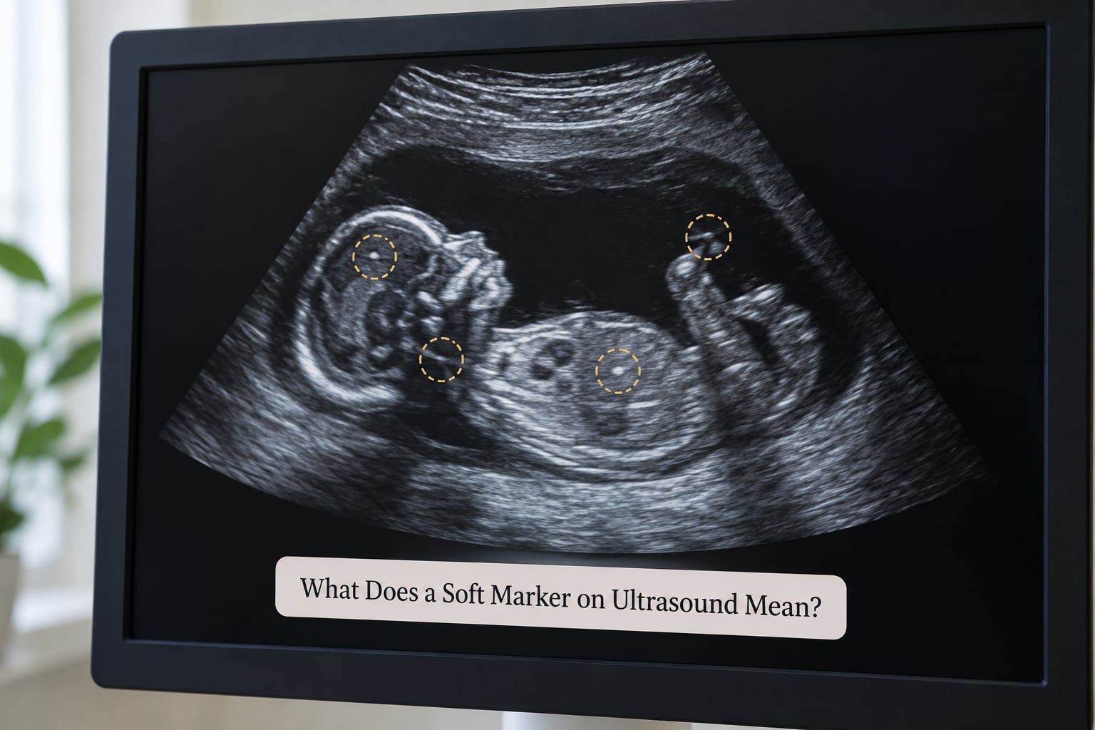
A soft marker is a minor anatomical variation seen on a prenatal ultrasound that is not a birth defect and does not affect your baby’s health or organ function. It is a nonspecific finding that may slightly adjust the statistical likelihood of a chromosomal condition, and most resolve before birth.
Two things make “soft” the important word. The finding is not an abnormality in the fetus’s structure or function, and it does not predict a condition on its own. It is a flag for a closer human look, nothing more.
Around 1 to 5 percent of detailed mid-pregnancy scans include at least one soft marker, with some clinics reporting 5 to 6 percent. The large majority of those pregnancies have entirely normal chromosomes and completely healthy babies.
If the phrase arrived on a report with no explanation, that is a gap in the conversation, not a signal about your baby. Ask what the specific marker is before you decide how much attention it deserves, because echogenic bowel and a hypoplastic nasal bone are very different conversations.
Why Do Sonographers Look for Soft Markers?
During the 18- to 22-week anatomy scan, the sonographer systematically checks specific features: the back of the neck, the nasal bone, the heart, the brain, the bowel and the kidneys. When one of those features falls slightly outside the typical range, it gets written down.
The reason to write it down is that these features are not equally common in pregnancies with and without aneuploidy. Statistically, they show up a little more often when a baby has an extra or missing chromosome. That difference is small, and it only becomes useful when combined with everything else known about the pregnancy.
Sonographers are not diagnosing. They describe what they see using a defined vocabulary, and a physician reads those descriptions in context. That context includes gestational age, how the baby was positioned during the scan, the quality of the images, and every other result on file.
Is a Soft Marker the Same as a Birth Defect?
No. A soft marker and a birth defect sit in different categories, and conflating them is the source of most scan-related panic.
A soft marker is a finding about how something looks, with no known effect on function. An anatomical finding describes the shape or structure of an organ. A confirmed structural anomaly, sometimes called a hard marker, is an actual abnormality in development that usually changes clinical management on its own, regardless of any genetic discussion.
Think of it as the difference between a finding and a diagnosis. A sonographer can see an echogenic intracardiac focus, a bright spot on a heart valve, and that bright spot turns out to be a small mineral deposit that many healthy adults also carry. It is a variation, not a defect.
| Category | What it is | What follows |
|---|---|---|
| Soft marker | Minor variation, normal structure, no effect on function | Weighed with age, screening and other findings |
| Anatomical finding | Describes the shape or structure of an organ | Judged by the specialist who examines it |
| Structural anomaly (hard marker) | A confirmed abnormality in development | Targeted imaging, MFM referral, plan of care |
What Do Common Soft Markers Mean?

These are the ones parents search for by name, because they are the ones most often written on the report without a plain-English note next to them.
Echogenic intracardiac focus (EIF)
Also called a bright spot or golf ball sign. It is a small calcium deposit on a heart valve, seen in a substantial share of healthy pregnancies and more often in boys. As an isolated finding it is widely treated as clinically insignificant, especially after a low-risk screen.
Choroid plexus cyst (CPC)
A fluid-filled pocket in the brain that makes fluid. CPCs usually resolve by the 24-week scan. They matter mainly when they persist, are large, or occur alongside other findings.
Echogenic bowel
Bowel that looks brighter than usual. This one gets more attention than most because it is linked to conditions that are not chromosomal at all, including blood swallowed in the womb, cystic fibrosis and congenital infection, which is why it often prompts closer monitoring rather than alarm.
Pyelectasis
Written as mild pyelectasis or pelviectasis. It means a small amount of urine is sitting in the kidney pelvis, and the kidneys are a touch wider than average. Very common, usually resolves, and one repeat scan is often all it gets.
Other markers you may see
Nuchal translucency or a thickened nuchal fold at the back of the neck, a hypoplastic nasal bone, a short femur or humerus, and a single umbilical artery are all described in a similar way. Septated hygroma and micrognathia are usually added to a list only when something else is already concerning.
| Finding | What it is | When assessed | Usual next step |
|---|---|---|---|
| Echogenic intracardiac focus | Calcium deposit on a heart valve | 18-22 week scan | Often none if isolated |
| Choroid plexus cyst | Fluid pocket in the brain | 18-22 week scan | Repeat scan to confirm resolution |
| Echogenic bowel | Brighter-than-usual bowel loops | 18-22 week scan | Targeted scan, sometimes monitoring |
| Pyelectasis | Mild widening of the kidney pelvis | 18-22 week scan | One repeat scan later in pregnancy |
| Nuchal translucency | Fluid at the back of the neck | 11-14 week scan | Depends on the measured value |
| Hypoplastic nasal bone | Small or unossified nasal bone | 11-14 week scan | Often combined with other screening |
| Single umbilical artery | One artery instead of two | 18-22 week scan | Growth monitoring later |
Does One Soft Marker Mean My Baby Has a Genetic Condition?
No. One marker does not mean that. It slightly adjusts a statistical estimate, and that estimate is built from several pieces of information rather than from the marker alone.
Clinicians weigh your age, any serum or cell-free DNA screening result, your family history, the measured size and appearance of the marker, how many were seen, and whether the rest of the anatomy looked normal. Isolated markers, particularly ones that resolve, carry very little weight in that calculation. International guidance, including the ISUOG and SMFM positions, holds that a single marker after a low-risk cell-free DNA result is generally not clinically significant.
On the other side of that, cell-free DNA is a screening test, not a diagnosis. A low-risk result lowers a chance; it does not erase it. Parents on r/NIPT raise this often, citing false-negative figures in the literature for conditions such as trisomy 18, sometimes up to about 7 percent. That is exactly the conversation to have with a genetic counselor, who can put the number in the context of your particular pregnancy rather than in a forum thread at midnight.
What Happens After a Soft Marker Is Found?
The steps vary with the marker and with how the rest of the pregnancy looks, but the usual sequence is short.
- Name the exact finding. Ask which marker it is, which side of the body, how large it measured, and whether it appeared on any earlier scan.
- Have the images rechecked. A senior sonographer or the reporting physician often reviews the pictures again, because positioning and image quality change what is visible. Positional artifact is a well-documented cause of findings that are not there on a repeat look.
- Time it correctly. Some markers are expected at 12 weeks and should not be judged at 20, and CPCs are usually re-scanned around 24 weeks to confirm they have gone.
- Decide on targeted imaging. A focused anatomy scan, or a fetal echocardiogram if a heart-related marker was noted, is often the whole plan.
- Ask about genetic counseling. A counselor reviews the full picture and explains screening versus diagnostic options such as amniocentesis or CVS, including what each would and would not tell you.
Many families stop at step three. Markers that look smaller on later scans, as one r/NIPT parent described after findings eased across three appointments, are a reassuring pattern rather than a worrying one.
How Is Risk Calculated From Multiple Soft Markers?
One marker and four markers are not the same conversation, and the difference matters more than most parents expect.
Clinicians do not simply count. They look at whether the findings cluster in one organ system, which often points to something local and structural, or across unrelated systems, which is the pattern that carries more weight for chromosomal considerations. Two markers in the same baby may also reflect one underlying variation rather than two independent ones.
When the number of markers is high, the usual response is not panic but a structured workup: a detailed or tertiary-level scan, cardiac imaging, genetic counseling and an honest conversation about whether diagnostic testing would change anything you would do with the pregnancy. Some markers carry more weight than others for specific conditions, and the specialist applying them is the person who knows which weight applies to yours.
Questions to Ask Your Doctor or Midwife
Prepare these before the appointment and bring a pen. Getting the finding named precisely is the single most useful thing you can do.
- Which specific marker was seen, and on which side of the body?
- Was it isolated, or were others found at the same time?
- Was it present on any earlier scan, and has it changed in size?
- Was the image reviewed by a senior sonographer or a radiologist?
- Does it affect my risk estimate at all, and by how much?
- Is a repeat scan or targeted imaging recommended, and when?
- Which of my results are screening and which are diagnostic?
- Would genetic counseling change anything about my care plan?
- Can I get the finding written in my records, in plain terms?
- When should I expect to hear back, and who do I call if I do not?
When to Seek Prompt Medical Advice
Contact your obstetric clinician or midwife to interpret a soft marker. Routine follow-up is the normal pace for this kind of finding.
Some symptoms need immediate attention, and they are about the pregnancy rather than the marker. Seek urgent care for heavy bleeding, severe or persistent abdominal pain, fluid leaking from the vagina, fever, regular contractions before 37 weeks, or a clear reduction in fetal movement. Your clinician will want to know about a marker you were told about, but these symptoms need attention whether or not a marker was ever mentioned.
Asking what a soft marker on ultrasound report means in writing also protects you later. Having the finding documented clearly means a second opinion, a change of provider or a future clinician is working from the same information you had.
Frequently Asked Questions
Are ultrasound soft markers always serious?
No. A soft marker is a minor variation, not a defect, and it does not affect how the baby’s organs work. Most appear in pregnancies with entirely normal chromosomes and resolve before birth. They are logged so a clinician can weigh them alongside your age and screening results. Serious ultrasound findings are called structural anomalies, and they trigger a different, faster pathway.
Does an isolated soft marker mean my baby has a chromosomal condition?
An isolated marker by itself does not mean a chromosomal condition. Guidance from ISUOG and SMFM holds that a single soft marker, particularly after a low-risk cell-free DNA result and with normal anatomy elsewhere, is generally not clinically significant. The estimate your clinician works from combines age, screening, family history and scan findings, so ask how your marker fits into that calculation rather than what it means alone.
Will I need another ultrasound if a soft marker is found?
Often, yes, though the repeat is usually routine rather than urgent. Choroid plexus cysts are commonly re-scanned around 24 weeks to confirm they have resolved, and pyelectasis often gets one later check. Your sonographer may also have the images re-reviewed before anything is scheduled, since positioning at the time of the scan affects what is visible. Ask for the specific reason and timeline rather than assuming.
Is increased nuchal translucency considered a soft marker?
It depends on the value. Nuchal translucency is measured at the 11- to 14-week scan and is used as part of first-trimester screening, so it is treated as a screening measurement rather than a single soft marker. A mildly increased measurement with normal blood tests is often reassuring, while a larger measurement or one alongside other findings usually leads to detailed imaging and genetic counseling. Ask how your number was interpreted.
Can a soft marker change the plan for my delivery?
Rarely on its own. An isolated marker with normal growth and normal anatomy does not usually affect where or how you give birth. Follow-up imaging may add a growth scan later in pregnancy, and some heart findings lead to a planned assessment after birth. If you are planning a home birth or a specific hospital, it is reasonable to mention the marker when you discuss your birth preferences.
Should I ask for genetic counseling after a soft-marker finding?
It is a reasonable request, especially with more than one marker, a family history, or a screen that was not conclusive. A genetic counselor reviews the full picture rather than the marker alone and explains what screening can and cannot rule out, plus what diagnostic testing would involve. Many parents find the session useful even when the outcome is reassurance, because it clarifies what happens next.
What to Do First
Ask one question at your next appointment: which specific marker was seen, and is it isolated. That single answer tells you more than hours of searching, and it is usually the point where the anxiety settles.
Soft markers are common, mostly temporary, and far more often a variation than a problem. What a soft marker on ultrasound means depends entirely on your own scan, your history and your clinician’s read of the whole picture, which is worth hearing from them directly rather than from a search result.