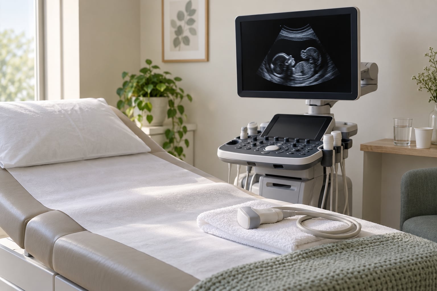
If you are searching for an anatomy scan at 20 weeks what they check, the short version is this: a sonographer systematically looks at your baby’s head and brain, face, spine, heart, abdomen, kidneys, limbs, genitals, placenta, umbilical cord, and the volume of amniotic fluid, and takes measurements along the way. It is a screening look at structure and growth. It is not a diagnosis, and a normal scan is the most common result. This guide walks through each area, what a measurement actually means, and what to ask afterward. It reflects current practice for 2026, and your own prenatal care team always has the final say for your pregnancy.
Table of Contents
- Anatomy Scan at 20 Weeks: What They Check
- Why the Scan Usually Happens Around 20 Weeks
- What an Anatomy Scan at 20 Weeks Can Show
- How the Baby’s Head, Face, Brain, and Spine Are Assessed
- How to Read the Measurement Percentiles on Your Report
- How the Heart Is Checked
- How the Abdomen, Kidneys, and Major Vessels Are Checked
- How the Bones, Limbs, and Genitals Are Assessed
- How the Placenta, Umbilical Cord, and Fluid Are Evaluated
- What the Scan Cannot Tell You
- How to Prepare and What to Ask at the Appointment
- When to Ask About Follow-Up or a Referral
- Frequently Asked Questions
- Is an anatomy scan at 20 weeks the same as a pregnancy ultrasound?
- How long does a 20-week anatomy scan usually take?
- Can the anatomy scan at 20 weeks confirm the baby’s sex?
- Can a 20-week anatomy scan detect Down syndrome?
- Do you still need a full bladder for a 20-week scan?
- What happens if the baby is measuring a little different from other measurements?
- What to Do Next
Anatomy Scan at 20 Weeks: What They Check

The mid-pregnancy ultrasound, also called the anatomy scan, anomaly scan, level 2 ultrasound, or 20-week ultrasound, is a detailed prenatal ultrasound usually offered between 18 and 22 weeks. The sonographer moves a handheld transducer over your abdomen, and sound waves build up a moving grayscale picture of the baby, the placenta, and the surrounding fluid.
Earlier scans answer dating and viability questions. The 20-week scan answers a different question: does the structure look typical? That is why it takes longer, why you will be repositioned several times, and why the appointment is booked for a block of 30 to 45 minutes rather than 10.
It is a screening assessment. A sonographer records what is seen and a clinician interprets the images in the context of your history, blood tests, and any earlier screening. A finding that needs explaining usually means more images, a repeat visit, or a referral, not a diagnosis on the spot. For decisions about your own pregnancy, speak with your obstetric clinician or midwife.
Why the Scan Usually Happens Around 20 Weeks
Gestational age is counted from the first day of your last menstrual period, or from an early ultrasound when the dates need firming up. By 20 weeks the baby is large enough for the bones to cast a clear outline, the head is proportionally smaller, and the fluid-filled spaces have settled into a pattern that makes comparison with standard measurements meaningful. Before about 18 weeks the spine and four-chamber heart view are harder to see through the small size of the chest.
After roughly 22 to 24 weeks the baby grows large, the bones ossify further, and the head often settles into the pelvis, which blocks the acoustic window for several views. So the window between 18 and 22 weeks exists mainly for imaging reasons rather than because something about your pregnancy is only safe then.
Scheduling moves for practical reasons. A first-trimester screening result that came back with a higher chance of a chromosome condition, a family history of a heart defect, diabetes, an increased chance of a twins pregnancy, or a previous pregnancy with an abnormality can all move the appointment earlier, often to 18 weeks, or later if growth measurements are better read at a bigger size. Two babies usually get a longer booking because the sonographer has to document each one separately.
What an Anatomy Scan at 20 Weeks Can Show
Ultrasound cannot see everything an X-ray or an MRI can, but at this gestational age it can show a surprising amount of structure. Here is the organ-by-organ list, which is also the closest thing to a checklist for the appointment itself.
| Body part | What is examined | What clinicians look for |
|---|---|---|
| Head and brain | Biparietal diameter, head circumference, nuchal fold, ventricles, cerebellum, midline structures | Skull shape, symmetry of the two hemispheres, fluid spaces, the small bone at the back of the skull, posterior fossa |
| Face | Facial profile, orbits, lips, palate, jaw | Symmetry, continuity of the lip line, a clear view of the palate, nasal bone |
| Spine | Vertebral column from neck to sacrum, spinal canal | Three parallel lines on cross-section, vertebrae counting down correctly, no separation of the backbone |
| Heart | Four-chamber view, both outflow tracts, rate and rhythm | Even chamber sizes, four distinct chambers, a fluid-filled stomach, valves moving through a full cycle |
| Chest and diaphragm | Lungs, diaphragm, chest shape | Symmetric chest, a smooth diaphragm, no bowel sitting high in the chest |
| Abdomen | Abdominal wall, stomach, bowel, liver, insertion of the cord | Wall closing at the umbilicus, a visible and filling stomach, bowel in the right place |
| Kidneys and bladder | Both kidneys, renal pelvis width, bladder | Two kidneys of similar appearance, a bladder that fills and empties across the visit |
| Spine and limbs | Femur and humerus length, arms, legs, hands, feet, joints | Three long bones in each limb, digits visible, joints that flex, no fixed angle at a joint |
| Genitals | External genital development | Appearance consistent with gestational age, if the angle allows |
| Placenta and fluid | Placenta location and grade, amniotic fluid index, umbilical cord with three vessels | Placenta on the back wall or low and away from the cervix, normal fluid volume, three cord vessels in cross-section |
How the Baby’s Head, Face, Brain, and Spine Are Assessed
The head gets three measurements that end up on your growth chart. Biparietal diameter measures across the widest part of the skull, head circumference wraps around it, and abdominal circumference measures around the belly. Femur length, the longest bone in the body, is usually added as a fourth. Four measurements is the usual set because one alone can be misleading, and comparing them with each other tells the clinician whether the baby is proportionate.
Inside the skull, the sonographer checks that the two fluid-filled ventricles are symmetrical, that the cerebellum and the space behind it are present, and that the corpus callosum, the band of tissue connecting the hemispheres, can be seen. Nuchal fold, the soft tissue thickness at the back of the neck, is measured in the first trimester, and if the baby is still in a position that allows it later, some clinicians take it again around this scan. A thicker-than-expected nuchal fold is one of the most commonly mentioned soft markers, and it is worth remembering that soft markers are findings, not diagnoses. Many of them disappear on later scans.
For the face, the sonographer works along the profile from the forehead to the chin, then checks the orbits, the lips, and the palate. The palate is the classic hard one, because an open mouth or a tongue pressed upward can hide it, and getting a clear view sometimes means the baby has to change position.
The spine is checked in two ways. Looking across the back gives the three-line appearance of a healthy cross-section. Then the sonographer counts vertebrae from the neck downward, because a mismatch there can signal a problem with the lower spine. Ultrasound does detect open neural tube defects such as spina bifida reasonably well at this age, but it is not a guarantee against every spinal cord difference.
How to Read the Measurement Percentiles on Your Report
Growth measurements come back as percentiles, not pass or fail marks. A value is compared with a reference range for babies of the same gestational age, and a percentile describes how many babies measure less or more. The middle of the curve is the 50th percentile, so roughly half of all healthy babies fall below it and half above it. A single measurement at the 20th or 80th percentile is ordinary. What gets attention is a measurement that is clearly out on its own, or a set of measurements that is consistently off in the same direction, or a change between scans that is faster than expected. Ask your clinician for the actual values and where they sit; “small” and “small for dates” mean different things to different people, and the numbers settle it.
How the Heart Is Checked
Most anatomy scans include a basic cardiac assessment rather than a full heart study. The sonographer captures a four-chamber view, the view that looks down on all four heart chambers at once, and checks that the chambers are proportionate and that the septum between left and right is intact. Both outflow tracts, the tubes leading out of the ventricles, are also recorded, along with the heart rate and rhythm over a period of time.
A useful indirect sign is a fluid-filled stomach. If the stomach has not filled normally, it can point to a problem with blood flow through the heart, so the sonographer often comes back to it partway through the appointment to check.
It is worth being straight about the limits here. A routine four-chamber view catches many significant defects, but it is not a screen for every heart condition. Some abnormalities only show up in views a standard scan does not acquire, which is why a dedicated fetal echocardiogram, a longer detailed study of the heart, is offered in some situations: a previous child or family member with a congenital heart defect, diabetes, certain chromosome screening results, a raised nuchal fold, or a heart anomaly already seen on the anatomy scan. Ask whether your risk factors call for one. And if something is flagged, remember the difference between a screening result and a diagnosis; the next step is usually a more detailed look, not a conclusion.
How the Abdomen, Kidneys, and Major Vessels Are Checked
The abdominal check starts with the wall, where the cord enters the body, because the skin and muscle should close neatly there. Then the sonographer looks for the stomach, which usually appears as a small dark pocket that fills and then empties over the course of the visit, and traces bowel gas in the lower abdomen. The diaphragm is checked as the boundary between chest and abdomen, to confirm that abdominal organs have not pushed up into the chest.
Both kidneys are assessed, and a measurement of the renal pelvis, the funnel where urine collects before it drains down the ureter, is often taken. A slightly wide renal pelvis, called pyelectasia, is one of the more frequently recorded soft markers at this scan. In most babies it resolves on its own, and the plan is usually to look again later rather than do anything now.
The cord itself gets a cross-section view. Two arteries and one vein, the three-vessel pattern, is what most babies have, and the umbilical arteries are also traced toward the bladder to see where they connect. The two major vessels running down the front of the abdomen, the aorta and the inferior vena cava, are checked for a normal number of vessels and for the position of the cord where it joins the placenta. A single umbilical artery changes how the rest of the pregnancy is monitored, so it is a specific thing to ask about if it is mentioned.
One note on the bladder: because the bladder fills slowly, it may not be easy to see at the start of the appointment. Sonographers often check it near the end, or ask you to come back after a walk.
How the Bones, Limbs, and Genitals Are Assessed
Limb review is partly a count and partly a length. The long bones in each arm and leg, femur, tibia, fibula, humerus, radius, and ulna, are all measured and counted, which is how a missing or shortened bone gets picked up. The hands and feet are checked for digits, the arms for symmetry, and the joints for movement. A fixed angle at a joint rather than one that flexes is a finding, and it is checked more than once, because babies hold unusual positions sometimes for no reason.
The general bone survey also covers the skull, ribs, and the small bones of the hands and feet, and the sonographer notes overall bone density. A shorter-than-expected long bone, or an arm or leg measuring notably shorter than its pair, gets measured a second time and often rechecked at a later visit, since positioning on one side can tighten the reading.
On genital development, the scan may show clear external anatomy. It is not, however, performed primarily to find out the sex, and a position where the legs are crossed or the cord is in the way can make the view impossible. Plenty of parents leave without an answer for exactly that reason. If your clinic does not share the sex, that is a policy and not a quality problem with the scan.
How the Placenta, Umbilical Cord, and Fluid Are Evaluated
The placenta is found and described by position and by how it attaches. Its location on the back wall is the most straightforward. A placenta on the front wall, which is called anterior, mostly adds glare and makes images harder to read, so the sonographer may approach from a different angle. A low-lying placenta, one whose edge sits within about 2 centimetres of the internal cervical opening, is measured precisely and usually rechecked later, because the placenta typically moves up as the uterus grows. The placenta is also graded, a way of describing how its appearance matures relative to the weeks of pregnancy.
Amniotic fluid is assessed as the amniotic fluid index, which is the sum of the deepest pocket of fluid measured in four areas around the baby. Too little or too much can matter for different reasons, and the clinician looks at the trend against previous visits rather than one number in isolation. An unusually large pocket, polyhydramnios, is often loosely linked to a baby that swallows less often, and low fluid, oligohydramnios, is associated with a baby that swallows more, or with leaking fluid, though the cause is worked out separately.
You will also notice the sonographer pausing to watch the baby’s chest and abdomen move, and sometimes tasting the fluid. None of it is a problem, and it is one of the quiet signs that things are working as they should.
What the Scan Cannot Tell You
It would be easy to read a clear picture on a screen as a guarantee of a healthy baby, and it is not one. Ultrasound depends on the position of the baby, the amount of fluid, the mother’s body size, and the bones in the way, so some views are genuinely unobtainable on the day.
Here is what the scan does not do. It cannot confirm or exclude a chromosome condition such as Down syndrome on its own; it may find features that raise or lower a statistical chance, and diagnostic answers come from blood tests such as NIPT, or from amniocentesis, which has to be discussed with your clinician because it carries its own small risks. It does not pick up every heart defect, and most soft markers are not conditions at all. It cannot rule out a functional or metabolic problem that leaves the anatomy looking normal, and some things are only picked up after birth, in the newborn examination or in a hearing and metabolic screen.
That is why the anatomy scan is one part of a pregnancy rather than a verdict on it. A normal result is reassuring and worth having. An incomplete or flagged result is a prompt for more information, not a conclusion, and the difference between those two sentences matters more than anything else you will read about scans online.
How to Prepare and What to Ask at the Appointment

Follow your clinic’s instructions first, because bladder and food advice differs between providers and some clinics have moved away from asking you to arrive with a full bladder at all. Many still ask you to drink water beforehand, because fluid in the bladder pushes the uterus up and gives the transducer something to work with. If yours does, the common advice is a couple of glasses of water in the hour before the appointment, and holding it rather than emptying it just before you walk in. Eat normally, since a scan that runs long is easier with something in your stomach, and wear a two-piece outfit so the gel does not reach a one-piece dress.
Allow a full hour in the building even though the scan itself often takes 30 to 45. A first visit is regularly longer, and clinics usually allow a longer slot for a first pregnancy, a first anatomy scan, or a twin pregnancy. Bring someone with you if you can. Many clinics allow a support person, and it is easier to have two people listening than to reconstruct the conversation afterwards. Bring written questions, because you will not remember them all in the room.
Expect a quiet room. Sonographers often work silently, concentrating on measurements, and that silence is not bad news. Some clinics will tell you results as they go, some will not say anything until the images are reviewed. Either style is normal, and asking at the start, will you tell me today or after the images are read, sets your expectations.
Questions that get useful answers include: are all the required views complete today, what measurements were taken and which percentile are they on, are there any soft markers, has the placenta moved away from the cervix, and is the amniotic fluid index within range. Add the ones specific to you: whether your risk factors call for a fetal echocardiogram, when the next growth scan is, and what the plan is if a view could not be obtained today.
On photos, ask before the visit. Some clinics print one or two images, some send a full gallery digitally, and some no longer print at all. Knowing in advance saves the disappointment of assuming a print is coming. Parents asking about saving and sharing images also ask about clinic policy, since recording video is restricted in a lot of departments, and about the apps people use to keep a private copy of their own images.
When to Ask About Follow-Up or a Referral
There are two different situations, and they are easy to mix up. The first is an incomplete scan: the baby was in a position where a view could not be obtained, or the sonographer could not get enough of a structure into a usable image. That is the most common follow-up, usually a repeat appointment in one to two weeks, often with advice to walk beforehand or to drink something cold to encourage movement. Plenty of people come back for a second visit and get a complete set of images with no other issue.
The second is a finding that needs more detail. That might mean a return appointment, a fetal echocardiogram for the heart, a more detailed growth measurement two to four weeks later, a maternal fetal medicine specialist, or a conversation with a genetic counselor. If your earlier screening showed a higher chance of a chromosome condition, ask whether an amniocentesis discussion belongs at this appointment rather than later. You are entitled to the time and information to make that decision yourself, and asking about the timeline is a reasonable question, not a difficult one to put.
Between appointments, contact your prenatal care team for anything urgent rather than waiting. Bleeding, fluid leaking, regular painful contractions, or a noticeable change in the baby’s usual movement pattern are reasons to call promptly. A mild one-off twinge in your own belly is different, and your clinician can tell you whether it is worth mentioning. For all decisions about your own pregnancy, your obstetric clinician, midwife, or maternal fetal medicine specialist is the right person; a guide like this can tell you what to ask, not what your baby needs.
Frequently Asked Questions
Is an anatomy scan at 20 weeks the same as a pregnancy ultrasound?
It is a type of ultrasound, and a more detailed one than a standard early scan. Ultrasounds early in pregnancy usually check dating, the heartbeat, and whether the pregnancy is progressing well. The 20-week anatomy scan is a level 2, or mid-pregnancy, scan focused on how the baby’s organs and skeleton are forming. It takes longer and records far more images, which is why it is booked as a longer appointment.
How long does a 20-week anatomy scan usually take?
Most appointments take about 20 to 45 minutes of scan time, and clinics usually block off closer to an hour for the visit. It runs longer when the baby is in a position where some views are hard to get, or when there is more to document, such as with twins or a higher-risk pregnancy. You may be asked to walk, roll onto your side, or come back after a drink to change the baby’s position.
Can the anatomy scan at 20 weeks confirm the baby’s sex?
It may show external genital development, but the scan is not performed mainly to determine sex, and the position of the baby can block a clear view entirely. Legs crossed, cord in the way, or a back turned toward the probe all make it difficult. Some clinics do not share the sex at this appointment by policy, so it is worth asking beforehand if you want to know and you have no preference about finding out.
Can a 20-week anatomy scan detect Down syndrome?
Not on its own, and it is not meant to. The scan looks at anatomy, and some chromosome conditions are associated with particular features such as a thicker nuchal fold or a shorter nasal bone. Those are called soft markers, and most are found in babies with typical chromosomes, so a marker adjusts a statistical estimate rather than settling anything. Answers come from blood tests such as NIPT, or from diagnostic testing such as amniocentesis, discussed with your clinician.
Do you still need a full bladder for a 20-week scan?
It depends on your clinic, and the answer has changed over the years. Many providers now ask you to arrive with a full bladder, or to drink water in the hour before the appointment, because fluid in the bladder lifts the uterus and gives clearer images. Others no longer require it, since improved equipment works well without. Follow whatever your clinic’s appointment instructions say, and ask them when the booking confirms, rather than relying on general advice.
What happens if the baby is measuring a little different from other measurements?
A single measurement that sits away from the middle is common and usually not a problem, because percentiles describe a spread rather than a pass mark. The sonographer records the values and the clinician interprets them against the due date, the other measurements, and any change from earlier scans. What matters is a set of measurements trending the same way, or one that is well outside the expected range. Ask for the numbers and where they sit on the chart.
What to Do Next
Turn up with your questions written down, a full bladder if your clinic asked for one, and someone beside you if you can. Listen to how your clinician explains anything that was flagged, and ask for the measurements in numbers so the percentages make sense later. Then leave with one clear next step: the date of the next growth scan, or a booked follow-up if any views were incomplete. Most people walk out with a normal report, a few photographs, and a due date that suddenly feels real.