How ultrasound dating works in early pregnancy is simpler than most first-time parents expect: a sonographer measures the length of the embryo from the top of its head to the bottom of its rump, the machine converts that millimetre figure into a gestational age, and your estimated due date is set 280 days from it. That measurement, called crown-rump length, is usually taken between 6 and 12 weeks.
This guide is general education about a common prenatal test, not advice about your own pregnancy. Your midwife, obstetrician or sonographer is the authority on what your scan means for you.
The short version:
- A dating scan does not measure your period. It measures the pregnancy itself.
- The key number is the crown-rump length, the head-to-rump distance of the embryo.
- The first-trimester scan sets the due date used for all later appointments.
- Later growth scans check development rather than re-dating the pregnancy.
Table of Contents
- What Is Ultrasound Dating in Early Pregnancy?
- How Is Gestational Age Measured on an Early Scan?
- What the sonographer looks for, in order
- How the crown-rump length becomes a gestational age
- Reading the abbreviations on your scan report
- How Does the Due Date Get Calculated?
- How Accurate Is an Early Pregnancy Ultrasound?
- Why Does the Due Date Sometimes Change?
- What Is the Difference Between Dating and Diagnostic Ultrasound?
- What Happens If the Pregnancy Is Too Small to Measure?
- How Ultrasound Dating Differs by Trimester
- Frequently Asked Questions
- Is an early pregnancy ultrasound more accurate than a due date calculated from the last menstrual period?
- Can an ultrasound tell me the exact day I conceived?
- Why was my due date changed after an early ultrasound?
- What does it mean if the gestational sac is too small to measure?
- Does ultrasound dating work the same way for twins or multiples?
- Conclusion
What Is Ultrasound Dating in Early Pregnancy?
A dating ultrasound is an early pregnancy scan, usually done between 6 and 12 weeks, that measures the size of the embryo to work out how many weeks pregnant you are and to set your due date. It is different from the date calculated from the last menstrual period, and different again from a conception date.
That difference matters. The last menstrual period method, sometimes called Naegele’s rule, assumes a 28-day cycle with ovulation on day 14. If your cycle runs 35 days, or you did not ovulate on schedule, that assumption puts the whole calculation weeks out.
Ultrasound dating skips the assumption. Instead of counting backwards from a period, it measures what has actually developed and counts forwards. For that reason, first-trimester ultrasound dating is considered the most accurate method available, and major guidelines from bodies such as NICE, ACOG, ISUOG and NHS pathways use it to establish gestational age when there is any uncertainty.
It is worth separating the vocabulary, because the terms get mixed up in appointment letters and online conversations:
- Gestational age — how far along the pregnancy is, counted from the first day of the last period. This is the number clinicians use.
- Embryonic or fetal age — time since conception, roughly two weeks less.
- Estimated date of delivery (EDD) — the calculated due date, sometimes written as EDD, EDC or EDD on your notes.
A scan estimates the first of those. It does not identify the exact day you conceived, and a scan in the first trimester does not screen for chromosomal conditions or structural problems.
How Is Gestational Age Measured on an Early Scan?
Before anything is measured, the sonographer checks where the pregnancy is and how it is developing. The structures appear in a predictable order, and each one tells the sonographer something about the stage of the pregnancy.
What the sonographer looks for, in order
- Gestational sac — the fluid-filled bubble that confirms the pregnancy has implanted inside the uterus. Usually visible from about 4½ to 5 weeks.
- Yolk sac — a small round structure that nourishes the embryo before the placenta takes over. Typically seen around 5 to 6 weeks.
- Fetal pole — the embryo itself, first appearing as a small bright line beside the yolk sac. Often visible from about 6 weeks.
- Cardiac activity — a flicker or pulse that confirms the heart has started beating. Often present by 6½ to 7 weeks.
Only once the fetal pole is measurable does dating become possible. Before that point the scan can confirm a pregnancy exists but cannot reliably tell you how far along it is.
How the crown-rump length becomes a gestational age
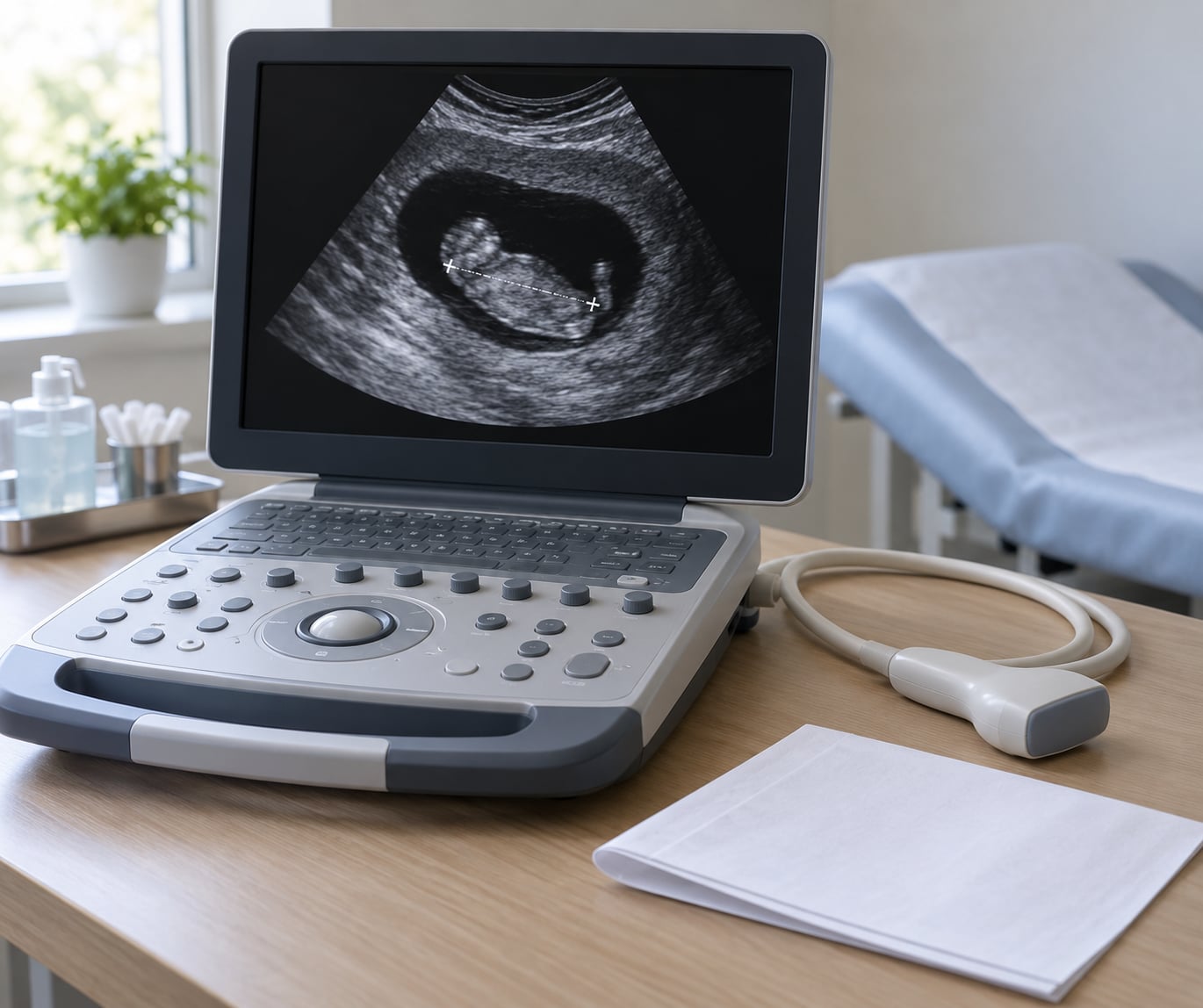
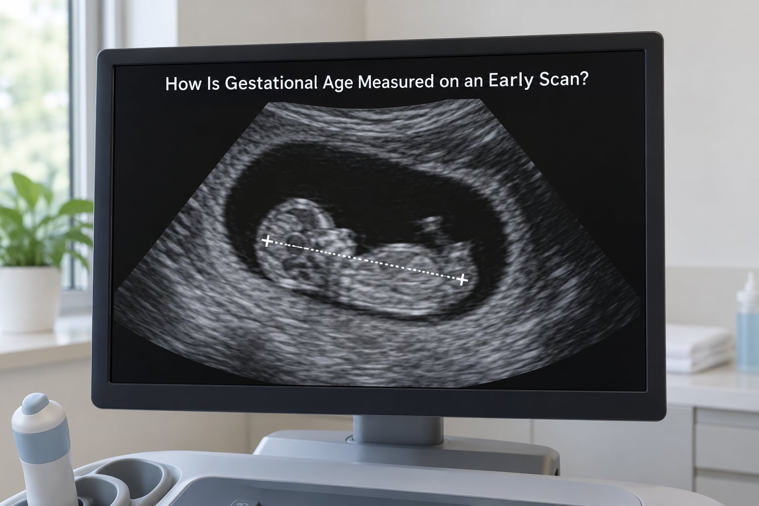
The probe sends high-frequency sound waves into the body and reads the echoes that bounce back off tissue boundaries. The sonographer moves the probe until the embryo appears in a clean, symmetrical position on screen, then places two electronic calipers: one at the top of the head, one at the bottom of the rump.
That span is the crown-rump length, or CRL. The embryo is measured lying flat rather than curled, because a curled position makes the number meaningless. Image quality is the other half of the job — if the head or rump is obscured by shadow, the sonographer will move the probe and measure again rather than accept a doubtful figure.
CRL is recorded in millimetres and matched against established reference tables. These are the widely used figures from the Robinson and Fleming charts, which underpin most first-trimester dating formulas:
| Gestational age | Typical CRL | What the scan usually shows |
|---|---|---|
| 6 weeks | 6 mm | Gestational sac, yolk sac, early fetal pole |
| 7 weeks | 10 mm | Fetal pole with cardiac activity |
| 8 weeks | 16 mm | Embryo with measurable proportions |
| 9 weeks | 23 mm | Distinguishable head, trunk and limb buds |
| 10 weeks | 32 mm | Clear limbs, cardiac activity steady |
| 11 weeks | 42 mm | Nuchal translucency can be assessed |
| 12 weeks | 54 mm | Approaching the end of first-trimester dating |
Reading the abbreviations on your scan report
Reports are written in a compressed shorthand. Learning four terms makes most early reports readable:
- GS — gestational sac, sometimes with a mean sac diameter in millimetres.
- CRL — crown-rump length, the dating measurement itself.
- FHR — fetal heart rate, usually reported in beats per minute, often seen between roughly 110 and 160 at this stage.
- NT — nuchal translucency, the fluid at the back of the neck measured at 11 to 14 weeks, not on the dating scan itself.
How Does the Due Date Get Calculated?
The conversion from millimetres to a date happens in four steps, and understanding them explains why a date can shift before it settles.
- Measure — the sonographer records the CRL in millimetres from a clear image.
- Convert — the machine or the reporting clinician looks up that measurement in a dating table, which returns a gestational age such as 8 weeks and 0 days.
- Project — the remaining days to term are calculated as 280 days minus the days already elapsed.
- Set the EDD — the scan date plus the remaining days gives the estimated date of delivery.
Put differently: EDD = scan date + (280 days − gestational age in days). If you are scanned on 4 June and the CRL reads 16 mm, that maps to 8 weeks and 0 days, so 224 days remain, and 4 June plus 224 days gives an estimated date of delivery of 14 January.
The resulting date is an estimate, and it is better understood as a range than a single day. A pregnancy that started on any one of about five days either side of the measured one would produce the same CRL figure, so a 40-week estimate carries an inherent spread of roughly a week before any measurement error is added.
Clinicians rarely quote the full range at the appointment because a number feels more useful than a range in the moment. But “how pregnant am I” means a window, not a timestamp, and the same window explains why two providers can quote slightly different dates for the same pregnancy without either being wrong.
How Accurate Is an Early Pregnancy Ultrasound?
First-trimester crown-rump dating is generally considered accurate to within about five days at 95% confidence when the pregnancy measures between roughly 8 and 13 weeks. Before 8 weeks the window widens, and dating scans in the 6-to-7-week range are commonly quoted as accurate to about a week either side.
Several factors affect precision:
- Where the pregnancy sits. An implantation lower in the uterus or at the side can distort the view and make a clean measurement harder to take.
- Imaging limitations. Body habit, uterine shape such as fibroids, or a full bladder that is needed for a transabdominal scan all change what is visible.
- Position of the embryo. A curled or oblique position gives an unreliable number, and experienced sonographers decline to record a measurement rather than guess.
- How early the scan happens. At 5½ weeks the embryo may simply be too small for a meaningful measurement.
None of this means the estimate is unreliable in practice. It means the date is an estimate. In forums, the consensus among parents is exactly that: the first-trimester scan is treated as the definitive date, and later scans are read as growth checks that run a week or two out in either direction without being a reason to re-date.
Why Does the Due Date Sometimes Change?
Because the ultrasound date is replacing an assumption, not correcting a mistake. The last-period method guesses when ovulation happened; the scan measures what developed. When the two disagree, the scan is the better evidence and the earlier date is set aside.
Cycle length explains most of the gap. Ovulation does not always fall on day 14 of a 28-day cycle, and later ovulation pushes conception later, which pushes the true gestational age back. An ultrasound reading that is smaller than the last-period calculation is therefore often the scan being right.
The same scan date with different measurements shows how the date moves. Here the scan happens on 4 June, and the last period began on 1 May, which by Naegele’s rule would predict 5 February:
| CRL measured | Gestational age | Days remaining | Estimated due date | Change from LMP date |
|---|---|---|---|---|
| 10 mm | 7 weeks 0 days | 231 | 21 January | About 2 weeks earlier |
| 16 mm | 8 weeks 0 days | 224 | 14 January | About 3 weeks earlier |
| 23 mm | 9 weeks 0 days | 217 | 7 January | About 4 weeks earlier |
| 32 mm | 10 weeks 0 days | 210 | 31 December | About 5 weeks earlier |
Small changes produce small moves: a few days of variation in embryo size shifts the due date by a comparable number of days. Larger shifts usually reflect a genuine difference between the assumed and actual conception date.
The source of most frustration is that two clinicians can appear to disagree. In practice the sonographer reports a measurement and the midwife or obstetrician sets the date from it, and the confusion usually comes from comparing a reported gestational age with a due date calculated by a different method. Asking which date was set from the scan, and on what measurement, resolves it faster than trying to work out the arithmetic yourself.
What Is the Difference Between Dating and Diagnostic Ultrasound?
A dating ultrasound answers one question: how far along is this pregnancy? A diagnostic ultrasound answers a different set of questions about anatomy, growth or a specific concern, and the two are often combined into a single appointment without being clearly labelled.
A dating scan looks for the gestational sac, the yolk sac, the fetal pole, cardiac activity, the number of embryos and the location of the pregnancy. A diagnostic scan looks at structure: the skull and brain, the spine, the heart chambers, the abdominal wall, the limbs, the kidneys, the placenta and the amniotic fluid.
The boundary is not about technology. Both use the same machine and often the same appointment. The difference is the question being asked and the measurements being recorded.
It also explains why a dating scan does not replace the routine anatomy scan at 18 to 22 weeks. The embryo is a few centimetres long at 8 weeks; there is not enough structure to assess. By the mid-second trimester the organs have developed enough to be examined properly, which is why that later scan exists as a separate appointment.
Nor does an early scan rule out every concern. It can confirm that a heartbeat is present and that development is on track for that size, which is genuinely reassuring, but conditions like chromosomal differences are detected through screening blood tests and the nuchal translucency measurement, and structural problems through the anatomy scan.
What Happens If the Pregnancy Is Too Small to Measure?
An early scan sometimes produces no usable number at all. The report may read “too small to measure” or “not assessable”, and the sonographer books a repeat scan rather than recording a guess.
Common reasons include:
- The pregnancy is genuinely earlier than estimated, which happens when ovulation occurred later than the last-period method assumes.
- The embryo is present but not yet positioned for a clean head-to-rump view, or the image quality is limited.
- Uterine anatomy such as fibroids or a tilted uterus makes the sac harder to see.
- The pregnancy is not in the usual location, and the sonographer needs more time and a different approach to examine the pelvis carefully.
When a measurement cannot be taken, the usual plan is a repeat scan after an interval of one to two weeks, by which time the embryo has grown enough to measure. Many people find that interval the hardest part, since the first scan is usually booked specifically because they want to know what is happening.
This is general information only. An inconclusive early scan is a clinical finding that should go to your own provider, who can interpret it alongside your history, any pain or bleeding, and your blood tests. Seek prompt advice rather than waiting for a repeat appointment if you have one-sided pain, bleeding or worsening symptoms.
How Ultrasound Dating Differs by Trimester
Dating is a first-trimester job, and later scans change what they are measuring rather than re-estimating the start date.
First trimester. The embryo grows at a tight, predictable rate, and its size maps closely onto gestational age. Crown-rump length works well from about 6 weeks to 13 weeks, which is exactly the window where a single small measurement gives the tightest estimate. This is why dating is locked in during this period.
Second trimester. Growth becomes more variable between individuals. Measurements such as biparietal diameter, head circumference, abdominal circumference and femur length are used to check that growth is proportionate, and nuchal translucency is measured at 11 to 14 weeks. A fetus measuring a week smaller at 20 weeks is ordinary variation, not a new due date.
Third trimester. Scans focus on position, placenta, amniotic fluid and estimated fetal weight. A one-off estimate late in pregnancy carries a wide margin, which is why clinicians prefer early measurements for dating and treat late growth scans as a single snapshot rather than a re-dating tool.
One regional difference is worth knowing. Some services book a dating scan at 6 to 10 weeks, while others, including many UK and New Zealand pathways, treat an 11-to-14-week scan as the first appointment and do the dating then. Both follow the same method; only the timing differs.
Frequently Asked Questions
Is an early pregnancy ultrasound more accurate than a due date calculated from the last menstrual period?
Yes, when the scan falls in the first trimester. The last-period method assumes a 28-day cycle with ovulation on day 14, so it drifts whenever a cycle is longer, shorter or irregular. An ultrasound measures the embryo directly and is generally accurate to within about five days when taken between 8 and 13 weeks, which is why guidelines use it when the two dates disagree.
Can an ultrasound tell me the exact day I conceived?
No. The scan converts a measurement in millimetres into a range of days rather than a single one, and an embryo of a given size could have started developing on any of several nearby days. An ultrasound date is also counted from the last menstrual period rather than from conception, so it runs roughly two weeks ahead of a conception date. For assisted conception, the clinic’s transfer date is usually kept instead.
Why was my due date changed after an early ultrasound?
Because the scan replaced an assumption with a measurement. Your previous date came from the last menstrual period, which assumes ovulation on day 14 of a 28-day cycle. The ultrasound found the pregnancy was a little smaller or larger than that estimate, so it produced a different gestational age. The first-trimester scan normally sets the final date, and later growth scans are not usually used to change it again.
What does it mean if the gestational sac is too small to measure?
It usually means the pregnancy is earlier than estimated, so the embryo has not yet grown into a position where a reliable crown-rump length can be taken. A small embryo for the calculated gestational age is common when ovulation happened later than the last-period method assumed. In most cases a repeat scan after one to two weeks produces a clear measurement. Your provider should interpret the finding for your own situation.
Does ultrasound dating work the same way for twins or multiples?
The method is the same, but the interpretation is not. The dating convention for a twin pregnancy depends on chorionicity, the type of twins, which is assessed early because it changes how the pregnancy is managed. Each embryo is measured individually, so their sizes are used to confirm they are developing consistently with each other and with the dating. Expect more time in the room and a more detailed report than for a singleton pregnancy.
Conclusion
How ultrasound dating works in early pregnancy comes down to one measurement and one calculation. A sonographer measures the crown-rump length, that number is matched to a gestational age, and 280 days are added to reach an estimated date of delivery. The date usually differs from a last-period calculation because the scan measures the pregnancy instead of assuming when conception happened, and once it is set in the first trimester it normally stays put.
Before you leave the appointment, it is worth asking what was measured, what gestational age it produced, and whether that date is now the one your appointments are scheduled against. Then take those numbers to your own midwife or obstetrician, who can tie the measurement to your history and your care plan. This article is general education and does not replace that conversation.